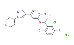
Crizotinib hydrochloride Datasheet DC Chemicals
Target Home > Products > Featured products
Cat.No DC22634
Name Crizotinib hydrochloride

Chemical Properties

CAS 1415560-69-8
Formula C21H23CL3FN5O
MW 486.797624826431
Storage 2 years -20°C Powder, 2 weeks 4°C in DMSO, 6 months -80°C in DMSO

Biological activity

Description CAS NO.:1415560-69-8
Product Name:Crizotinib hydrochloride
Synonyms:Crizotinib hydrochloride;3-[(1R)-1-(2,6-dichloro-3-fluorophenyl)ethoxy]-5-(1-piperidin-4-ylpyrazol-4-yl)pyridin-2-amine,hydrochloride;CS-1156
EINEC:
Molecular Formula:C21H23CL3FN5O
Molecular Weight:486.797624826431
Target: IC50: 11 nM (c-Met), 24 nM (ALK)
In Vivo PF-2341066 reveals the ability to cause marked regression of large established tumors (> 600 mm3) in both the 50 mg/kg/day and 75 mg/kg/day treatment cohorts, with a 60% decrease in mean tumor volume over the 43-day administration schedule in the GTL-16 model. In an another study, PF-2341066 displays the ability to completely inhibits GTL-16 tumor growth for >3 months, with only 1 of 12 mice exhibiting a significant increase in tumor growth over the 3-month treatment schedule at 50 mg/kg/day. A significant dose-dependent reduction of CD31-positive endothelial cells is observed at 12.5 mg/kg/day, 25 mg/kg/day, and 50 mg/kg/day in GTL-16 tumors, indicating that inhibition of MVD shows a dose-dependent correlation to antitumor efficacy. PF-2341066 displays a significant dose-dependent reduction of human VEGFA and IL-8 plasma levels in both the GTL-16 and U87MG models. Marked inhibition of phosphorylated c-Met, Akt, Erk, PLCλ1, and STAT5 levels is observed in GTL-16 tumors following p.o. administration of PF-2341066[1]. PF-2341066 prevents osteosarcoma behavior associated with primary tumor growth as well as metastasis. In nude mice treated with PF-2341066 via oral gavage, the growth and associated osteolysis and extracortical bone matrix formation of osteosarcoma xenografts are prevented by PF-2341066[3]. Treatment of c-MET-amplified GTL-16 xenografts with 50 mg/kg PF-2341066 elicits tumor regression that is associated with a slow reduction in 18F-FDG uptake and decreases expression of the glucose transporter 1, GLUT-1[4].
In Vitro PF-2341066 displays similar potency against c-Met phosphorylation in mIMCD3 mouse or MDCK canine epithelial cells with IC50 of 5 nM and 20 nM, respectivly. PF-2341066 shows improved or similar activity against NIH3T3 cells engineered to express c-Met ATP-binding site mutants V1092I or H1094R or the P-loop mutant M1250T with IC50 of 19 nM, 2 nM and 15 nM, respectively, compared with NIH3T3 cells expressing wild-type receptor with IC50 of 13 nM. In contrast, a marked shift in potency of PF-2341066 is observed against cells engineered to express c-Met activation loop mutants Y1230C and Y1235D with IC50 of 127 nM and 92 nM, respectively, compared with wild-type receptor. PF-2341066 also potently prevents the phosphorylation of c-Met in NCI-H69 and HOP92 cells, with IC50 of 13 nM and 16 nM, respectively, which express the endogenous c-Met variants R988C and T1010I, respectively[1]. PF-2341066 also potently inhibits NPM-ALK phosphorylation in Karpas299 or SU-DHL-1 ALCL cells with an IC50 of 24 nM. PF-2341066 potently prevents cell proliferation, which is associated with G(1)-S-phase cell cycle arrest and induction of apoptosis in ALK-positive ALCL cells with IC50 of 30 nM, but not ALK-negative lymphoma cells[2]. Besides, PF-2341066 prevents osteosarcoma behavior associated with primary tumor growth (i.e., proliferation and survival) as well as metastasis[3].
Kinase Assay Cells are seeded in 96-well plates in media supplemented with 10% fetal bovine serum (FBS) and transferred to serum-free media [with 0.04% bovine serum albumin (BSA)] after 24 h. In experiments investigating ligand-dependent RTK phosphorylation, corresponding growth factors are added for up to 20 min. After incubation of cells with PF-2341066 for 1 h and/or appropriate ligands for the designated times, cells are washed once with HBSS supplemented with 1 mM Na3VO4, and protein lysates are generated from cells. Subsequently, phosphorylation of selected protein kinases is assessed by a sandwich ELISA method using specific capture antibodies used to coat 96-well plates and a detection antibody specific for phosphorylated tyrosine residues. Antibody-coated plates are (a) incubated in the presence of protein lysates at 4°C overnight; (b) washed seven times in 1% Tween 20 in PBS; (c) incubated in a horseradish peroxidase-conjugated anti-total-phosphotyrosine (PY-20) antibody (1:500) for 30 min; (d) washed seven times again; (e) incubated in 3,3′,5,5′-tetramethyl benzidine peroxidase substrate to initiate a colorimetric reaction that is stopped by adding 0.09 N H2SO4; and (f) measured for absorbance in 450 nm using a spectrophotometer.
Cell Assay Tumor cells are seeded in 96-well plates at low density in media supplemented with 10% FBS (growth media) and transferred to serum-free media (0% FBS and 0.04% BSA) after 24 h. Appropriate controls or designated concentrations of PF-2341066 are added to each well, and cells are incubated for 24 to 72 h. Human umbilical vascular endothelial cells (HUVEC) are seeded in 96-well plates in EGM2 media for 5 to 6 h at > 20,000 cells per well and transferred to serum-free media overnight. The following day, appropriate controls or designated concentrations of PF-2341066 are added to each well, and after 1 h incubation, HGF is added to designated wells at 100 ng/mL. A 3-(4,5-dimethylthiazol-2-yl)-2,5-diphenyltetrazolium bromide assay is done to determine the relative tumor cell or HUVEC numbers.
Animal Administration Athymic mice bearing xenografts (300-800 mm3) are given PF-2341066 in water by oral gavage at designated dose levels. At designated times following PF-2341066 administration, mice are humanely euthanized, and tumors are resected. Tumors are snap frozen and pulverized using a liquid nitrogen-cooled cryomortar and pestle, protein lysates are generated, and protein concentrations are determined using a BSA assay. The level of total and phosphorylated protein is determined using a capture ELISA or immunoprecipitation-immunoblotting method.

References

[1]. Zou HY, et al. An orally available small-molecule inhibitor of c-Met, PF-2341066, exhibits cytoreductive antitumor efficacy through antiproliferative and antiangiogenic mechanisms. Cancer Res. 2007, 67(9), 4408-4417. [2]. Christensen JG, et al. Cytoreductive antitumor activity of PF-2341066, a novel inhibitor of anaplastic lymphoma kinase and c-Met, in experimental models of anaplastic large-cell lymphoma. Mol Cancer Ther. 2007, 6(12 Pt 1), 3314-3322. [3]. Sampson ER, et al. The orally bioavailable met inhibitor PF-2341066 inhibits osteosarcoma growth and osteolysis/matrix production in a xenograft model. J Bone Miner Res. 2011, 26(6), 1283-1294. [4]. Cullinane C, et al. Differential (18)F-FDG and 3'-deoxy-3'-(18)F-fluorothymidine PET responses to pharmacologic inhibition of the c-MET receptor in preclinical tumor models. J Nucl Med. 2011 Aug;52(8):1261-7
Return Policy
If you are in any way unsatisfied with your purchase, you may return any item(s) within 365 days of its original purchase date.
Please provide your Order Number in the email. We strive to reply to all email inquiries within one business day.
Tel: +86-21-58447131
Fax: +86-21-61642470

Email:
sales@dcchemicals.com
order@dcchemicals.com

Website:
www.dcchemicals.com