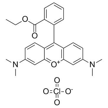
TMRE Datasheet DC Chemicals
Target Home > Products > Featured products
Cat.No DC65474
Name TMRE

Chemical Properties

CAS 115532-52-0
Formula C26H27CLN2O7
MW 514.9548
Storage 2 years -20°C Powder, 2 weeks 4°C in DMSO, 6 months -80°C in DMSO

Biological activity

Description CAS NO.:115532-52-0
Product Name:Tetramethylrhodamine ethyl ester perchlorate
Synonyms:3,6-Bis(dimethylamino)-9-(2-(ethoxycarbonyl)phenyl)xanthylium perchlorate;[6-(dimethylamino)-9-(2-ethoxycarbonylphenyl)xanthen-3-ylidene]-dimethylazanium,perchlorate;3,6-Bis(dimethylamino)-9-(2-(ethoxycarbonyl)-phenyl)xanthylium;Tetramethylrhodamine ethyl ester perchlorate;Tmre;TMRE [Tetramethylrhodamine, ethyl ester, perchlorate];RB3096;TMRE perchlorate;AK117804;Xanthylium, 3,6-bis(dimethylamino)-9-[2-(ethoxycarbonyl)phenyl]-, perchlorate (1:1);AM62669;ST24041271;3,6-bis(dimethylamino)-9-[2-(ethoxycarbonyl)phenyl]-xanthylium perchlorate;Tetramethyl;Q27147928;Tetramethylrhodamine ethyl ester
EINEC:
Molecular Formula:C26H27CLN2O7
Molecular Weight:514.9548
Target:
In Vivo
In Vitro TMRE is a mitochondria specific dye (λex=550 nm, λem=575 nm)[1]. Multidirectional dynamic movement of TMRE is observed in epithelial cells and bidirectional dynamic movement is seen in the superficial cortical fiber cells of live bovine lenses. In the epithelium, the movement of TMRE fluorescence is up to 5 μm/min whereas in the superficial cortex the observed movement is up to 18.5 μm/min. The movement of TMRE fluorescence is abolished with treatment of the uncoupler, carbonyl cyanide m-chlorophenylhydrazone (CCCP)[2].
Kinase Assay
Cell Assay The entire experiment should be performed at room temperature because temperature will directly impact mitochondrial transmembrane potential and TMRE staining. Cells should never be placed, centrifuged, incubated, or washed at 4°C or have ice-cold buffers or media added. Treat the cells with a cytotoxic stimulus. Harvest cells and resuspend at 5×10⁵ cells/mL in culture medium containing 150 nM TMRE. Incubate for 5 min at room temperature in the dark. Add Carbonyl cyanide 4-(trifluoromethoxy)phenylhydrazone (FCCP) (5 μM final concentration) to an aliquot of untreated cells and incubate for 5 min at room temperature in the dark. Turn on the appropriate laser on the flow cytometer. Set up a histogram plot to detect TMRE using log scale[1].
Animal Administration

References

[1]. Crowley LC, et al. Measuring Mitochondrial Transmembrane Potential by TMRE Staining. Cold Spring Harb Protoc. 2016 Dec 1;2016(12):pdb.prot087361. [2]. Bantseev V, et al. Confocal laser scanning microscopy imaging of dynamic TMRE movement in the mitochondria of epithelial and superficial cortical fiber cells of bovine lenses. Mol Vis. 2005 Jul 14;11:518-23.
Return Policy
If you are in any way unsatisfied with your purchase, you may return any item(s) within 365 days of its original purchase date.
Please provide your Order Number in the email. We strive to reply to all email inquiries within one business day.
Tel: +86-21-58447131
Fax: +86-21-61642470

Email:
sales@dcchemicals.com
order@dcchemicals.com

Website:
www.dcchemicals.com