| Description: |
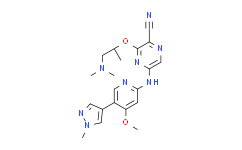
CCT244747 is a potent, orally bioavailable and highly selective CHK1 inhibitor, with an IC50 of 7.7 nM; CCT244747 also abrogates G2 checkpoint with an IC50 of 29 nM. |
| In Vivo: |
CCT244747 (100 mg/kg po, qd7d) significantly reduces tumor burden in human tumor xenografts. CCT244747 (100-300 mg/kg, po) inhibits gemcitabine-induced pS296 CHK1 for up to 24 h in HT29 colon tumor xenografts[1]. CCT244747 (75 mg/kg, p.o.) in combination with gemcitabine has potent antitumor effects in HT29 colon tumor xenografts and Calu6 human lung cancer xenografts. CCT244747 (150 mg/kg p.o.) also shows antitumor activities with irinotecan in SW620 human colon tumor xenografts[2]. CCT244747 (100 mg/kg, p.o.) exhibits radiosensitization activity in Cal27 xenografts[3]. |
| In Vitro: |
CCT244747 poorly inhibits CHK2 (IC50 >10 μM) and CDK1 (IC50 >10 μM). CCT244747 has potent activities against CHK1, RSK1, RSK2, AMPK, BRSK1, IRAK1,and TrkA, with >80% inhibition. CCT244747 (10 μM) exhibits <25% inhibition of the other ion channels including hNav1.5, hKv4.3/hKChIP2, hCav1.2, hKv1.5, hKCNQ1/hminK, hHCN4[1]. CCT244747 inhibits FLT3 with an IC50 of 600 nM. CCT244747 (0.5 μM) overcomes genotoxic-induced S and G2 cell cycle arrest in human colon cancer cell lines. CCT244747 inhibits cellular CHK1 function with IC50s ranging from 29 nM to 170 nM for cellular G2 checkpoint abrogation (MIA, mitosis induction assay) in HT29, SW620, MiaPaCa-2, and Calu6 cell lines; the GI50s are between 0.33 and 3μM. CCT244747 (0.3 μM) inhibits SN38 and gemcitabine-induced CHK1 activity in HT29 and SW620 colon cancer cell lines and this correlates with abrogation of cell cycle arrest, induction of DNA damage and apoptosis[2]. CCT244747 (0.5-2.0 μM) increases the sensitivity of bladder and head and neck cancer cell lines (T24, RT112 and Cal27) to radiation[3]. |
| Kinase Assay: |
CHK1 kinase activity is measured in a microfluidic assay that monitored the separation of a phosphorylated product from its substrate. The assay is run on an EZ Reader II using separation buffer containing CR-8 (500 nM). An ECHO® 550 acoustic dispenser is used to generate duplicate 8 pt dilution curves directly into 384 polypropylene assay plates. For each compound a 50 μM stock concentration in 100% DMSO is used. The total amount of DMSO dispensed per well is 250 nL to give a final assay concentration of 2.5% DMSO and compound concentrations in the range 0.5-1000 nM. To this assay plate, 6 PL CHK1 (2 nM final concentration, in-house protein preparation), 2 PL peptide 10 (5-FAM-KKKVSRSGLYRSPSMPENLNRPR-COOH, 1.5 PM final concentration) and 2 PL ATP (90 PM final concentration) all diluted in kinase buffer (HEPES 50 mM, NaN3 0.02%, BSA 0.01%, sodium orthovanadate 0.1 mM, DTT 1 mM, MgCl2 2 mM, Tween20 0.1%) are added. The plate is sealed and centrifuged (1 min, 1000 rpm) before ncubation for 1 h at room temperature. The reaction is stopped by the addition of separation buffer (90 PL). The plate is read on an EZ Reader II, using a 12-sipper chip with instrument settings of -1.5 psi and 1750 ΔV. The percentage conversion of product from substrate is generated automatically and the percentage inhibition is calculated relative to blank wells (containing no enzyme and 2.5% DMSO) and total wells (containing all reagents and 2.5% DMSO). IC50 values are calculated in GraphPad Prism5 using a non linear regression fit of the log (inhibitor) vs response with variable slope equation[1]. |
| Cell Assay: |
Compound cytotoxicity and the ability of CHK1 inhibitors to enhance SN38 (the active metabolite of the topoisomerase I inhibitor irinotecan) and gemcitabine (an antimetabolite) cytotoxicity is assessed using a 96 h sulforhodamine B assay (SRB). HT29 or SW620 cells are seeded at 1.6 to 3.2 × 10³ cells per well in 96-well plates in a volume of 160 μL medium and allowed to attach for 36 h prior to treatment. For cytotoxicity assays, CHK1 inhibitors (10 mM stock in DMSO) are serially diluted in medium from a starting concentration of 250 PM and then 40 PL is added to appropriate wells in quadruplicate to give a final concentration range of 50-0.1 PM (10 concentrations). Genotoxic agents (SN38; 10 mM stock in DMSO) are serially diluted in medium from a starting concentration of 2 PM and 40 PL is added to each well inquadruplicate to give final concentrations from 200-0.39 nM (10 concentrations). Cells are incubated for 96 h (four doublings) at 37°C in a humidified 5% CO2 environment and then fixed and stained with SRB. Appropriate controls are included and results are expressed as the concentration of test compound required to inhibit cell growth by 50% relative to untreated controls (SRB IC50). Potentiation assays involved adding a fixed SRB IC50 concentration of either gemcitabine or SN38 in a volume of 20 μL of medium (10× final concentration), to each well in quadruplicate and mixing for 1 min. CHK1 inhibitor (10 mM stock) is serially diluted from a starting concentration of 50 PM in medium and 20 PL is added per well in quadruplicate to give a final concentration range of 5-0.039 PM (8 concentrations). After mixing for 1 min the cells are incubated at 37°C in a humidified atmosphere for 96 h (four doublings) prior to fixing and SRB staining. Untreated and genotoxic alone treated controls are included and results are expressed as the concentration of CHK1 inhibitor required to inhibit cell growth by 50% (potentiation IC50). The potentiation index (PI) is used as a measure of the ability of the CHK1 inhibitor to enhance SN38 or gemcitabine cytotoxicity and is the ratio of the SRB IC50 versus potentiation IC50 (PI = SRB IC50 / Potentiation IC50)[1]. |
| Animal Administration: |
Female BALB/c mice (6 weeks old) are kept in a controlled environment with food and sterilized water available ad libitum. Animals weighed 20 (±2) g at the time of experiment. Dosing solutions are prepared by dissolving the compounds in 10% DMSO and 5% Tween20 in 85% saline. The compounds are administered i.v. and p.o., individually. Animals are warmed before receiving a single i.v. bolus injection into a lateral tail vein. Oral administration is by oral gavage. Blood is collected at selected time points (1 h and 6 h after dosing) by cardiac puncture under anesthesia into heparinized syringes, transferred to micro centrifuge tubes, and centrifuged at 4500 × g for 2 min to obtain plasma. Quantitative analysis is performed by high performance liquid chromatography tandem mass spectrometry on a triple quadrupole instrument using multiple reaction monitoring of selected transitions with olomoucine used as internal standard. Quantitation is performed against a standard curve ranging from concentrations of 2-1000 nM in the matrix measured. Quality controls are included at the level of 25, 250 and 750 nM. If required, samples are diluted in the matrix of interest[1]. |
| References: |
[1]. Lainchbury M, et al. Discovery of 3-alkoxyamino-5-(pyridin-2-ylamino)pyrazine-2-carbonitriles as selective, orally bioavailable CHK1 inhibitors. J Med Chem. 2012 Nov 26;55(22):10229-40.
[2]. Walton MI, et al. CCT244747 is a novel potent and selective CHK1 inhibitor with oral efficacy alone and in combination with genotoxic anticancer drugs. Clin Cancer Res. 2012 Oct 15;18(20):5650-61.
[3]. Patel R, et al. An orally bioavailable Chk1 inhibitor, CCT244747, sensitizes bladder and head and neck cancer cell lines to radiation. Radiother Oncol. 2017 Mar;122(3):470-475. |

 To enhance service speed and avoid tariff delays, we've opened a US warehouse. All US orders ship directly from our US facility.


