| Description: |
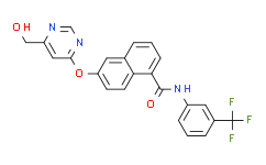
BFH772 is a potent oral VEGFR2 inhibitor, which is highly effective at targeting VEGFR2 kinase with an IC50 value of 3 nM. |
| In Vivo: |
BFH772 at 3 mg/kg orally dosed once per day potently inhibits melanoma growth (by 54-90% for primary tumor and 71-96% for metastasis growth) as depicted by treatment to control ratios. Dose–response curves of BFH772 at 0.3, 1, and 3 mg/kg demonstrate that even at the lowest concentrations, this naphthalene-1-carboxamide inhibits VEGF induced tissue weight and TIE-2 levels but only reaches statistical significance at 1 mg/kg and above[1]. |
| In Vitro: |
BFH772 is highly selective; apart from inhibiting VEGFR2 at 3 nM IC50, it also targets B-RAF, RET, and TIE-2, albeit with at least 40-fold lower potency. BFH772 is inactive (IC50>10 μM; >2 μM for cKIT) against all other tyrosine specific- and serine/threonine-specific protein kinases tested. BFH772 inhibits VEGFR2 with IC50 of 4.6±0.6 nM in CHO cells. BFH772 inhibits VEGFR2 with IC50 of 3 nM in HUVEC cells. BFH772 inhibits the ligand induced autophosphorylation of RET, PDGFR, and KIT kinases, with IC50 values ranging between 30 and 160 nM. BFH772 is selective (IC50 values >0.5 μM) against the kinases of EGFR, ERBB2, INS-R, and IGF-1R and against the cytoplasmic BCR-ABL kinase. IC50 of BFH772 (<0.01 nM, n=2) demonstrates that they abrogated VEGF induced proliferation at remarkably low nM concentrations[1]. |
| Kinase Assay: |
In vitro kinase assay is based on a filter binding assay, using the recombinant GST-fused kinase domains expressed in baculovirus and purified over glutathione-sepharose, γ-[33P]ATP as the phosphate donor, and poly(Glu:Tyr 4:1) peptide as the acceptor. Each GST-fused kinase is incubated under optimized buffer conditions [20 mM Tris-HCl buffer (pH 7.5), 1-3 mM MnCl2, 3-10 mM MgCl2, 3-8 μg/mL poly(Glu:Tyr 4:1), 0.25 mg/mL polyethylene glycol 20000, 8 μM ATP, 10 μM sodium vanadate, 1 mM DTT] and 0.2 μCi γ-33P ATP in a total volume of 30 μL in the presence or absence of a test substance for 10 min at ambient temperature. The reaction is stopped by adding 10 mL of 250 mM EDTA. Using a 384-well filter system, half the volume is transferred onto an Immobilon-polyvinylidene difluoride membrane. The membrane is then washed extensively and dried, and scintillation counting is performed. IC50s for compounds are calculated by linear regression analysis of the percentage inhibition[1]. |
| Cell Assay: |
Different Ba/F3 cell lines rendered IL-3 independent by transduction with various constitutively active tyrosine kinases are grown in RPMI 1640 medium containing 10% fetal calf serum. For maintenance of parental Ba/F3 cells, the medium is additionally supplemented with 10 ng/mL interleukin-3 (IL-3). For proliferation assays, Ba/F3 cells are seeded on 96-well plates in triplicates at 10000 cells per well and incubated with various concentrations of compounds for 72 h followed by quantification of viable cells using a resazurin sodium salt dye reduction readout (commercially known as Alamar Blue assay). IC50s are determined with the XLFit Excel Add-In using a four-parameter dose response model[1]. |
| Animal Administration: |
Mice[1] Female FVB mice weighing between 18 and 20 g are housed in groups of six. Porous chambers containing VEGF (2 μg/mL) in 0.5 mL of 0.8% w/v agar (containing heparin, 20 U/mL) are implanted subcutaneously in the flank of the mice (n=6 per group). VEGF induces the growth of vascularized tissue around the chamber. This response is dose-dependent and can be quantified by measuring the weight and TIE-2 levels of the tissue. Mice are treated either orally once daily with compounds or vehicle (PEG200 100%, 5 mL/kg) starting 4-6 h before implantation of the chambers and continuing for 4 days. The animals are sacrificed for measurement of the vascularized tissues 24 h after the last dose. Tissue weight is taken and then a lysate prepared for TIE-2 ELISA analysis . Rats[1] Catheters are implanted into the femoral artery and vein of naïve female rats strain OFA for BFH772, and BAW2881, or in the jugular vein and femoral artery in female Sprague-Dawley rats for compounds 4, 9, and 10. Animals are allowed to recover for 96 h and are housed in single cages with free access to food and water throughout the experiment. Female OFA rats received 2.5 mg/kg of BAW2881 dissolved in ethanol/dimethylisosorbide/polyethylene glycol400/D5W (10/15/35/40 v/v) or 1 mg/kg of BFH772 dissolved in N-methyl pyrrolidone/polyethylene glycol200 (30:70, v/v) via injection into the femoral vein. D5W is glucose 5%/water (v/v). Oral administration: BAW2881 and BFH772 are formulated as a micronized suspension (dissolved/suspended in 0.5% carboxymethyl cellulose in distilled water) and administered by gavage to female OFA rats to deliver a dose of 25 mg/kg for BAW2881 or 3 mg/kg BFH772 (n=4 rats per group). For compounds 4, 9, and 10, female Sprague-Dawley rats at 8 weeks of age received an intravenous dose of 3 mg/kg 4, 9, and 10, formulated in ethanol/NMP/polyethylene glycol400/D5W (10/10/50/30) (n=2 rats per group), or a suspension in 0.5% carboxymethyl cellulose in distilled water dosed at 50 mg/kg (n=3 rats per group). At the allotted times, blood samples are collected into heparinized tubes, and the amount of compound in plasma determined by HPLC/MS-MS. |
| References: |
[1]. Bold G, et al. A Novel Potent Oral Series of VEGFR2 Inhibitors Abrogate Tumor Growth by Inhibiting Angiogenesis. J Med Chem. 2016 Jan 14;59(1):132-46. |


