| Description: |
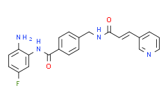
Chidamide is a synthetic benzamide-type HDAC inhibitor, inhibits HDAC1, HDAC2, HDAC3 and HDAC10 with IC50s of 95, 160, 67 and 78 nM, respectively. |
| In Vivo: |
Inhibition of tumor growth by Chidamide (CS055) treatment is observed in a dose-dependent manner, demonstrating the anti-tumor activity of Chidamide. Control tumors grow to an average volume of 14.51 cm3 after 20 days, and Chidamide-treated tumors grow to 11.68, 11.05 and 8.45 cm3, corresponding to 19.54%, 23.83% and 41.80% growth inhibition respectively. The average tumor mass in animals treated with vehicle is 9.4±2.7 g and is 8.4±2.4 g for animals treated with low-dose Chidamide. In animals treated with a moderate dose of Chidamide, tumor mass is 7.6±1.6 g and those receiving high-dose Chidamide has a tumor mass of 5.4±1.5 g (P<0.01). Additionally, Chidamide treatment prolongs the survival of nude mice bearing HL60 xenografts. Moreover, the level of lipid peroxidation product (MDA), which is a presumptive measure of ROS-mediated injury, is increased in tumor tissue accompanied by treatment of Chidamide, suggesting that Chidamide-induced ROS generation might play a role[2]. |
| In Vitro: |
Chidamide (CS055) at low concentrations dramatically inhibits cell proliferation in each cell line. After Chidamide treatment, cells arrest at the G0/G1 phase in a dose-dependent manner. Western blot analysis indicats that cyclin E1 and E2 protein expression is down-regulated after Chidamide treatment, which is consistent with the cell-cycle analysis. As the changes in cyclin E1 are much more significant than cyclin E2, cyclin E1 is up-regulated in HL60 and K562 cells by lentiviral transduction. The effect on leukaemia proliferation by Chidamide inhibition are largely weakened when cyclin E1 is overexpressed. It is therefore likely that cyclin E1 levels are decreased by Chidamide which induces cell-cycle arrest at the G0/G1 phase[2]. Chidamide causes a significant concentration-dependent inhibitory effect on cell proliferation in comparison to the vehicle-treated cells (P<0.05). The maximal inhibitory effect is reached at 5 μM[3]. |


