 To enhance service speed and avoid tariff delays, we've opened a US warehouse. All US orders ship directly from our US facility.


| Cas No.: | 2097381-85-4 |
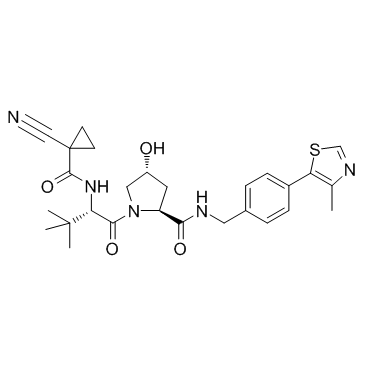
| Chemical Name: | (2S,4R)-1-((S)-2-(1-Cyanocyclopropanecarboxamido)-3,3-dimethylbutanoyl)-4-hydroxy-N-(4-(4-methylthiazol-5-yl)benzyl)pyrrolidine-2-carboxamide |
| Synonyms: | VH298;VH298 |
| SMILES: | C(C1(C(NC(C(C)(C)C)C(N2CC(O)CC2C(NCC2C=CC(C3SC=NC=3C)=CC=2)=O)=O)=O)CC1)#N |
| Formula: | C27H33N5O4S |
| M.Wt: | 523.652 |
| Purity: | >98% |
| Sotrage: | 2 years -20°C Powder, 2 weeks 4°C in DMSO, 6 months -80°C in DMSO |
| Publication: | [1]. Frost J, et al. Potent and selective chemical probe of hypoxic signalling downstream of HIF-α hydroxylation via VHL inhibition. Nat Commun. 2016 Nov 4;7:13312. |
| Description: | VH-298 is a highly potent inhibitor of the VHL:HIF-α interaction with a Kd value of 80 to 90 nM, used in PROTAC technology. |
| Target: | Kd: 80 to 90 nM (VHL:HIF-α)[1] |
| In Vitro: | VH-298 is a potent, cell permeable and non-toxic chemical probe that triggers the hypoxic response by blocking the VHL. VH-298 is a highly potent inhibitor of the VHL:HIF-α interaction with Kd values of 90 and 80 nM in isothermal titration calorimetry and competitive fluorescence polarization assay. VH-298 binds with VHL complex very fast and dissociates slowly. VH-298 at 50 μM concentration exhibits negligible off-target effects in vitro against more than 100 tested cellular kinases, GPCRs and ion channels. VH-298 is cell permeable and not toxic to cells. The measured permeability of VH-298 is found to be 19.4 nm s -1. VH-298 induces concentration- and time-dependent on-target specific accumulation of hydroxylated HIF-α in human cell lines, including HeLa cancer cells and renal cell carcinoma 4 (RCC4) cells. VH-298 increases mRNA levels of EPO by 2.5-fold in RCC4-HA-VHL, but not in VHL-null RCC4-HA, indicating that pharmacological inhibition of VHL is able to stimulate endogenous EPO synthesis. VH-298 proves as effective as hypoxia in raising PHD2 and HK2 protein levels, however in HFF the BNIP3 protein level increases more with VH-298 treatment than hypoxia treatment[1]. |
| Kinase Assay: | VH-298 is screened at 50 μM concentration against a panel of 50 kinases. The remaining kinase activity is recorded in the end of the assay. The data is reported as average % activity remaining of assay duplicates for each kinase tested[1]. |
| Cell Assay: | Death of CTLs is analyzed by staining with 4′,6-diamidino-2-phenylindole (DAPI). Cells are plated in 96-well plates at 1×10⁶ and treated with VHL inhibitors (VH-298) and respective non-binding cis-analogues for 24 h. Cells are spun down and resuspended in HBSS containing DAPI to identify dead and dying populations[1]. |
| References: | [1]. Frost J, et al. Potent and selective chemical probe of hypoxic signalling downstream of HIF-α hydroxylation via VHL inhibition. Nat Commun. 2016 Nov 4;7:13312. |