| Description: |
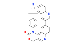
ETP-46464 is an effective mTOR and ATR inhibitor with IC50s of 0.6 and 14 nM, respectively. |
| In Vitro: |
ETP-46464 (ATRi) also inhibits DNA-PK, PI3Kα and ATM with IC50s of 36 nM, 170 nM and 545 nM, respectively[1]. Platinum-sensitive and -resistant ovarian, endometrial and cervical cancer cell lines are treated with varying levels of Cisplatin (0-50 µM) with or without the ETP-46464 (5.0 µM) and/or the KU55933 (10.0 µM) for 72 h. Single-agent dose response analyses of ETP-46464 and KU55933 in a subset of cell lines reveal a wide LD50 range of 10.0±8.7 and 38.3±7.6 µM respectively. Co-treatment doses are chosen based on these studies and previously published evidence of phospho-Chk1 (Ser345) and phospho-ATM (Ser1981) inhibition following ionizing radiation exposure and dose response treatments with ETP-46464 and KU55933. Treatment with ETP-46464 significantly increases the response of Cisplatin in all cell lines tested, resulting in 52-89% enhancement in activity and are synergistic. The combined inhibition of ATR and ATM enhances the response of Cisplatin to a level equivalent to that observed using ETP-46464 alone. These effects are independent of p53 status, and are observed in all gynecologic (GYN) cancer cells tested. Treatment with ETP-46464, but not KU55933, not only sensitizes these GYN cancer cell lines to Cisplatin, but also enhances the response of Carboplatin[2]. |
| Kinase Assay: |
Compounds (e.g., ETP-46464) and control inhibitors are delivered directly to cell media (100 μL per well) with a multi-well pipette at a final concentration of 10 μM. Media content is homogenized by carefully vortexing plates at 500 rpm. Prior to 4-hydroxy-tamoxifen (4-OHT) addition, Compounds (e.g., ETP-46464 ) are incubated at 37ºC for 15 minutes. Next, to induce ATR activity, 4-OHT is added to all wells and incubated for 60 minutes at 37ºC. Finally, cells are fixed with paraformaldehyde and processed for IF. Every compound (e.g., ETP-46464) is analyzed at least in three independent experiments[1]. |
| Cell Assay: |
Cells are trypsinized with 0.25% Trypsin-EDTA and counted with 0.4% Trypan Blue using an automated cell counter and plated in 96-well plates at 5000 cells per well for KLE, HEC1B and HELA and 10,000 cells per well for OVCAR3, A2780, A2780-CP20 and SIHA. After cells attach and reach approximately 60% confluency (24-48 h post seeding), media is removed and replaced with fresh media containing Cisplatin (0, 0.78, 1.56, 3.13, 6.25, 12.5, 25 or 50 µM) or Carboplatin (0, 1.56, 3.13, 6.25, 12.5, 25, 50 or 100 µM) in 0.15% DMSO, 5 µM ETP-46464, 10 µM KU55933, or a combination of 5 µM ETP-46464 and 10 µM KU55933 and incubated for 72 h. Final concentrations of ETP-46464 and KU55933 utilized are based on prior evidence indicating inhibition of ATR and ATM signaling, respectively. Single-agent dose response analyses of ETP-46464 and KU55933 in a subset of cell lines revealed a wide LD50 range (10.0±8.7 and 38.3±7.6 µM, respectively). Similarly, cells are treated with fresh media containing Cisplatin (0, 0.78, 1.56, 3.13, 6.25, 12.5, 25 or 50 µM) in 0.08% DMSO and 5 µM VE-821. Cell viability is assessed using the MTS CellTiter 96 Aqueous One Solution Cell Proliferation Assay. After a 2 h incubation at 37°C, absorbance is measured at 490 nm using a microplate spectrophotometer. Three biological replicates are performed for each cell line where each inhibitor(s)/Cisplatin concentration is assayed in triplicate for each experiment[2]. |
| References: |
[1]. Toledo LI, et al. A cell-based screen identifies ATR inhibitors with synthetic lethal properties for cancer-associated mutations. Nat Struct Mol Biol. 2011 Jun;18(6):721-7.
[2]. Teng PN, et al. Pharmacologic inhibition of ATR and ATM offers clinically important distinctions to enhancing platinum or radiation response in ovarian, endometrial, and cervical cancer cells. Gynecol Oncol. 2015 Mar;136(3):554-61. |


