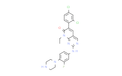
| Description: |
FRAX486 is a p21-activated kinase (PAK) inhibitor with IC50s of 14, 33 and 39 nM for PAK1, PAK2 and PAK3, respectively. |
| Target: |
PAK1:14 nM (IC50)
PAK2:33 nM (IC50)
PAK3:39 nM (IC50) |
| In Vivo: |
FRAX486 displays the highest penetrance of blood–brain barrier in DISC1-knockdown C57BL/6 mice. Daily administration of FRAX486, but not that of vehicle, between P35 and P60 blocks the exacerbated spine loss during adolescence. In addition to the significant blockade of spine elimination, a trend of enhanced spine generation is observed by treatment with FRAX486. FRAX486 treatment ameliorates a deficit in prepulse inhibition in adulthood[2]. |
| In Vitro: |
In vitro kinase assays using pure enzymes reveal IC50s for FRAX486 between 10-100 nM for PAK1-3, while the IC50 of 779 nM for PAK4 is just below the micromolar range. For FRAX486, an EC50 value of 500 nM has been reported from cells (5-50 fold higher than IC50). FRAX486 (30 μM) inhibits endothelin-1 and -2 induced contractions. In WPMY-1 cells, FRAX486 (24 h) induces concentration-dependent (1-10 μM) degeneration of actin filaments. This is paralleled by attenuation of proliferation rate, being observed from 1 to 10 μM FRAX486. Cytotoxicity of FRAX486 in WPMY-1 cells is time- and concentration-dependent. FRAX486 significantly reduces the relative proliferation rate in the remaining populations of WPMY-1 cells. While 68% of solvent-treated (24 h) cells shows proliferation, proliferation rate after application of FRAX486 (1-10 μM, 24 h) ranges around 45%. FRAX486 (1-10 μM, 24 h) causes concentration-dependent degeneration of actin filaments. Actin filaments in solvent-treated control cells are arranged to bundles, forming long and thin protrusions, with elongations from adjacent cells overlapping each other. FRAX486 in concentrations of 1 μM causes partial loss of actin organization, including regressing degree of actin polymerization and degeneration of protrusions. FRAX486 in concentrations of 5 or 10 μM causes complete breakdown of filament organization, resulting in a rounded cell shape without protrusions[1]. |
| Cell Assay: |
WPMY-1 cells are plated with a density of 50,000/well on a 16-well chambered coverslip. After 24 h, cells are treated with FRAX486 (1, 5, 10 μM), IPA3 (1, 5, 10 μM), or DMSO. After further 24 h, the medium is changed to a 10 mM 5-ethynyl-2’-deoxyuridine (EdU) solution in FCS-free medium containing inhibitors or solvent. 20 h later, cells were fixed with 3.7% formaldehyde. EdU incorporation is determined using the “EdU-Click 555” cell proliferation assay. In this assay, incorporation of EdU into DNA is assessed by detection with fluorescing 5-carboxytetramethylrhodamine (5-TAMRA). Counterstaining of all nuclei is performed with DAPI. Cells are analyzed by fluorescence microscopy (excitation: 546 nm; emission: 479 nm)[1]. |
| Animal Administration: |
Mice[2] The fasted male C57BL/6 mice are used. For FRAX486, i.v. dose is 3 mg/kg using a 1 mg/mL solution in 20% (wt/vol) 2-hydroxypropyl-β-cyclodextrin in water, and per oral administration (o.s.) (PO) dose is 30 mg/kg in a 3 mg/mL solution in water. For the in vivo experiment, FRAX486 is intraperitoneally administered [10 μg/BW (g)] once daily from P35 to P60, which provides brain levels at >175 nM. |
| References: |
[1]. Wang Y, et al. P21-Activated Kinase Inhibitors FRAX486 and IPA3: Inhibition of Prostate Stromal Cell Growth and Effects on Smooth Muscle Contraction in the Human Prostate. PLoS One. 2016 Apr 12;11(4):e0153312.
[2]. Hayashi-Takagi A, et al. PAKs inhibitors ameliorate schizophrenia-associated dendritic spine deterioration in vitro and in vivo during late adolescence. Proc Natl Acad Sci U S A. 2014 Apr 29;111(17):6461-6. |


