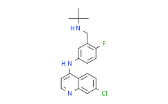
| Description: |
Niraparib hydrochloride (MK-4827 hydrochloride) is an excellent PARP1 and PARP2 inhibitor with IC50 of 3.8 and 2.1 nM, respectively. |
| In Vivo: |
Niraparib is well tolerated and demonstrates efficacy as a single agent in a xenograft model of BRCA-1 deficient cancer. Niraparib is well tolerated in vivo and demonstrates efficacy as a single agent in a xenograft model of BRCA-1 deficient cancer. Niraparib is characterized by acceptable pharmacokinetics in rats with plasma clearance of 28 (mL/min)/kg, very high volume of distribution (Vdss=6.9 L/kg), long terminal half-life (t1/2=3.4 h), and excellent bioavailability, F = 65%[1]. Niraparib enhances radiation response of p53 mutant Calu-6 tumor in both cases, with the single daily dose of 50 mg/kg being more effective than 25 mg/kg given twice daily[3]. |
| In Vitro: |
Niraparib inhibits PARP activity with EC50=4 nM and EC90=45 nM in a whole cell assay. Niraparib inhibits proliferation of cancer cells with mutant BRCA-1 and BRCA-2 with CC50 in the 10−100 nM range. Niraparib displays excellent PARP 1 and 2 inhibition with IC50=3.8 and 2.1 nM, respectively, and in a whole cell assay[1]. To validate that Niraparib inhibits PARP in these cell lines, A549 and H1299 cells are treated with 1 μM Niraparib for various times and measured PARP enzymatic activity using a chemiluminescent assay. The results show that Niraparib inhibits PARP within 15 minutes of treatment reaching about 85% inhibition in the A549 cells at 1 h and about 55% inhibition at 1 h for the H1299 cells[2]. |
| Kinase Assay: |
Enzyme assay is conducted in buffer containing 25 mM Tris, pH 8.0, 1 mM DTT, 1 mM spermine, 50 mM KCl, 0.01% Nonidet P-40, and 1 mM MgCl2. PARP reaction contains 0.1 μCi [3H]NAD+ (200 000 DPM), 1.5 μM NAD+, 150 nM biotinylated NAD+, 1 μg/mL activated calf thymus, and 1−5 nM PARP-1. Autoreactions utilizing SPA bead-based detection are carried out in 50 μL volumes in white 96-well plates. Compounds (e.g., Niraparib) are prepared in 11-point serial dilution in 96-well plate, 5 μL/well in 5% DMSO/H2O (10× concentrated). Reactions are initiated by adding first 35 μL of PARP-1 enzyme in buffer and incubating for 5 min at room temperature and then 10 μL of NAD+ and DNA substrate mixture. After 3 h at room temperature, these reactions are terminated by the addition of 50 μL of streptavidin-SPA beads (2.5 mg/mL in 200 mM EDTA, pH 8). After 5 min, they are counted using a TopCount microplate scintillation counter. IC50 data is determined from inhibition curves at various substrate concentrations[1]. |
| Cell Assay: |
The inhibition of PARP is analyzed in A549 and H1299 cells using the HT Universal Chemiluminescent PARP Assay Kit. Briefly, cells are treated with DMSO or 1 μM niraparib for 15, 30, 60, or 120 minutes, trypsinized, and transferred to a pre-chilled tube. The cells are washed twice with ice cold PBS and resuspended in cold PARP extraction buffer. The cell suspensions are incubated on ice for 30 minutes with periodic vortexing to disrupt the cell membrane. The suspensions are centrifuged and the supernatant transferred to a pre-chilled tube on ice. The histone coated wells of the 96-well plate are rehydrated with 1X PARP buffer and incubated at room temperature for 30 minutes. The PARP buffer is removed and 20 μg of protein as determined by the Bio-Rad Protein Assay is added to each well followed by diluted PARP-HSA enzyme and 1X PARP buffer. The strip wells are then incubated at room temperature for 60 minutes, washed twice with PBS containing 0.1% Triton X-100, and then washed with PBS. Diluted Strep-HRP is added to the strip wells and incubated for 60 minutes at room temperature. The wells are washed again as before. Equal volumes of PeroxyGlow A and B are combined and added to the wells and chemiluminescent readings are obtained immediately using a plate-reader[2]. |
| Animal Administration: |
Mice[3] Female nude mice (Ncr Nu/Nu) are randomly assigned to treatment groups consisting of 5 to 8 mice each when tumors grew to 6.0 mm in diameter at which time treatment with Niraparib is initiated. Niraparib is given at a dose of 25 mg/kg twice daily or 50 mg/kg once daily for either 21 days or is discontinued at 9 days from the time tumors reached 8 mm in diameter. Fractionated local tumor irradiation (XRT) is given when tumors reach 8.0 mm in diameter (7.7-8.2 mm). Radiation (2 Gy per fraction) is delivered to the tumor-bearing leg of mice once daily for 14 consecutive days or twice daily for 7 consecutive days using a small-animal irradiator consisting of two parallel-opposed 137Cs sources, at a dose rate of 5 Gy/min. During irradiation un-anesthetized mice are mechanically immobilized in a jig so that the tumor is centered within a 3.0 cm diameter radiation field and the animal’s body shielded from radiation exposure. On the day when both Niraparib and radiation are given, drug is administered 1 h before the first radiation dose of the day. |
| References: |
[1]. Jones P, et al. Discovery of 2-{4-[(3S)-piperidin-3-yl]phenyl}-2H-indazole-7-carboxamide (MK-4827): a novel oral poly(ADP-ribose)polymerase (PARP) inhibitor efficacious in BRCA-1 and -2 mutant tumors. J Med Chem. 2009 Nov 26;52(22):7170-85.
[2]. Bridges KA, et al. Niraparib (MK-4827), a novel poly(ADP-Ribose) polymerase inhibitor, radiosensitizes human lung and breast cancer cells. Oncotarget. 2014 Jul 15;5(13):5076-86.
[3]. Wang L, et al. MK-4827, a PARP-1/-2 inhibitor, strongly enhances response of human lung and breast cancer xenografts to radiation. Invest New Drugs. 2012 Dec;30(6):2113-20. |


