| Description: |
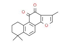
Tanshinone IIA (Tan IIA) is one of the main fat-soluble compositions in the root of red-rooted salvia. Tanshinone IIA may suppress angiogenesis by targeting the protein kinase domains of VEGF/VEGFR2. |
| Target: |
VEGF/VEGFR2[1] |
| In Vitro: |
The anti-tumor effect of Tanshinone IIA includes inhibiting tumor cell proliferation, disturbing tumor cell cycle, promoting tumor cell apoptosis, and inhibiting tumor cell invasion and transfer. Tanshinone IIA has anti-proliferative effects on A549 cells: the IC50 of Tanshinone IIA after 24, 48 and 72 h are 145.3, 30.95 and 11.49 μM, respectively. The CCK-8 assay is used to evaluate the proliferative activity of A549 cells treated with Tanshinone IIA (2.5-80 μM) for 24, 48 and 72 h, respectively. The CCK-8 results show that Tanshinone IIA can significantly inhibit A549 cell proliferation in a dose- and time-dependent manner. Obvious apoptosis and cell growth inhibition of A549 cells are observed after drug treatment for 48 h (concentrations used are approximately IC50 values: Tanshinone IIA 31 μM on A549). Western blotting finds that 48 h exposures to Tanshinone IIA (31 μM) in A549 cells, downregulates expression of VEGF and VEGFR2 protein in both drug treatment groups vs. vehicle[1]. Tanshinone IIA, one of the most abundant constituents of the root of Salvia miltiorrhiza, protects rat myocardium-derived H9C2 cells against apoptosis. Treatment of H9C2 cells with Tanshinone IIA inhibits angiotensin II-induced apoptosis by downregulating the expression of PTEN (phosphatase and tensin homolog), a tumor suppressor that plays a critical role in apoptosis. Tanshinone IIA inhibits angiotensin II (AngII)-induced apoptosis by downregulating the expression of phosphatase and tensin homolog (PTEN)[2]. Tanshinone IIA decreases the protein expression of EGFR, and IGFR blocking the PI3K/Akt/mTOR pathway in gastric carcinoma AGS cells[3]. |
| Cell Assay: |
A549 cells are counted in logarithmic phase and 6000 cells (90 μL volume) are placed in 96-well plates. 10 μL varying concentrations of Tanshinone IIA (final concentrations 80, 60, 40, 30, 20, 15, 10, 5 and 2.5 μM) and ADM (final concentrations 8, 4, 2, 1, 0.5 and 0.25 μM) are added into drug groups, while negative control group (vehicle group) is only added 10 μL DMSO or normal saline without Tanshinone IIA or ADM. Cells are incubated for an additional 2 h with CCK-8 reagent (100 μL/mL medium) and the absorbance is read at 450 nm using a microplate reader. Cell proliferation inhibition rates are calculated according to the following formula: the proliferation inhibition ratio (%)=1-[(A1-A4)/(A2-A3)]×100, where, A1 is the OD value of drug experimental group, A2 is the OD value of blank control group, A3 is the OD value of the RPMI1640 medium without cells, and A4 is the OD value of drugs with the same concentration as A1 but without cells. The IC50 value, which represents the concentration of the drug that demonstrates 50% of cell growth inhibition, is calculated by nonlinear regression analysis using GraphPad Prism software[1]. |
| References: |
[1]. Xie J, et al. The antitumor effect of tanshinone IIA on anti-proliferation and decreasing VEGF/VEGFR2 expression on the human non-small cell lung cancer A549 cell line. Acta Pharm Sin B. 2015 Nov;5(6):554-63.
[2]. Zhang Z, et al. Tanshinone IIA inhibits apoptosis in the myocardium by inducing microRNA-152-3p expression and thereby downregulating PTEN. Am J Transl Res. 2016 Jul 15;8(7):3124-32.
[3]. Su CC, et al. Tanshinone IIA decreases the protein expression of EGFR, and IGFR blocking the PI3K/Akt/mTOR pathway in gastric carcinoma AGS cells both in vitro and in vivo. Oncol Rep. 2016 Aug;36(2):1173-9. |


