 To enhance service speed and avoid tariff delays, we've opened a US warehouse. All US orders ship directly from our US facility.


| Cas No.: | 1001600-56-1 |
| Chemical Name: | BV-6 |
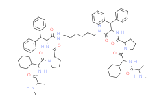
| Synonyms: | BV-6;BV 6;Dianemycin, monosodium salt;4,4'-(1,6-Hexanediyl)bis[N-methyl-L-alanyl-(2S)-2-cyclohexylglycyl-L-prolyl-beta-phenyl-L-phenylalaninamide;BV6;N,N'-(hexane-1,6-diyl)bis(1-{(2S)-2-cyclohexyl-2-[(N-methyl-L-alanyl)amino]acetyl}-L-prolyl-beta-phenyl-L-phenylalaninamide):trifluoroacetic acid(1:x) |
| SMILES: | O=C(NCCCCCCNC([C@@H](NC([C@H](CCC1)N1C([C@H](C2CCCCC2)NC([C@H](C)NC)=O)=O)=O)C(C3=CC=CC=C3)C4=CC=CC=C4)=O)[C@@H](NC([C@H](CCC5)N5C([C@H](C6CCCCC6)NC([C@H](C)NC)=O)=O)=O)C(C7=CC=CC=C7)C8=CC=CC=C8 |
| Formula: | C70H96N10O8 |
| M.Wt: | 1205.5734577179 |
| Purity: | >98% |
| Sotrage: | 2 years -20°C Powder, 2 weeks 4°C in DMSO, 6 months -80°C in DMSO |
| Description: | BV6 is an antagonist of cIAP1 and XIAP, members of the inhibitors of apoptosis (IAP) family. |
| In Vivo: | Murine cIAP-1, cIAP-2 and XIAP expressions are clearly observed in the cytoplasm of both epithelial and stromal cells of implants, whereas Survivin is mainly expressed in the nuclei BV6 treatment for 4 weeks attenuated the intensity of IAPs expression. The size of lesions range from ~2 to 7 mm in diameter. The monolayer epithelial cell lining of the cyst is shown. After immunohistochemical staining, cytokeratin and vimentin are positively stained, whereas calretinin is negative. After BV6 treatment for 4 weeks, the total number of lesions (4.6 versus 2.8/mouse), the average weight (78.1 versus 32.0 mg/mouse) and the surface area (44.5 versus 24.6 mm2/mouse) of lesions are significantly less than in the controls. In the endometrial gland epithelia or stroma, the percentage of Ki67-positive cells decreases after BV6 treatment[2]. |
| In Vitro: | HCC193 has an IC50 of 7.2 μM in MTS assays, while H460 cells are not reduced to 50% viability even with 30 μM BV6 treatment. Administration of 1 μM BV6 to HCC193 cells induces complete depletion of cIAP1 levels at 1 hour post-treatment, while a decrease in XIAP levels is not seen until 24 hours following addition of drug. Similarly, 5 μM BV6 fully depletes cIAP1 at 1 hour and begin to reduce XIAP at 24 hours in H460 cells. In parallel findings, cIAP1 levels are decreased in response to a small dose of 0.25 μM BV6 in both cell lines, whereas trace amounts of XIAP are still present at 5μM BV6. HCC193 cells demonstrates noticeable cleaved caspase-3 levels beginning 12 hours post-incubation with 1μM BV6, and cleaved caspase-3 levels continued to increase in a time-dependent manner over 48 hours. Treatment of HCC193 cells with 1 μM BV6 for 24 hours causes a significant survival curve shift in HCC193 cells relative to DMSO-treated cells, with a DER=1.38 (p<0.05)[1]. BV6 (2 and 5 µM) significantly represses BrdU incorporation in ectopic and eutopic (disease-free and myomas) ESCs. An ~30% decrease of BrdU incorporation is observed in both groups after treatment with 5 µM BV6[2]. |
| Cell Assay: | H460 and HCC193 cell lines are cultured in RPMI-1640 supplemented with 10% fetal bovine serum and 1% penicillin/streptomycin. Cell viability is measured using the CellTiter 96 Aqueous Non-Radioactive Cell Proliferation Assay kit. 5000 cells/well are seeded into 96-well plates in triplicate. Following adhesion of cells to the wells, increasing concentrations of BV6 are added into different wells. Control groups are exposed to the same concentration of DMSO. The final concentrations of 333 μg/mL MTS and 25 μM PMS are added to each well 24 hours later. After two hours incubation at 37°C in humidified 5% CO2, plates are read at the absorbance of 490 nm on a microplate reader. Relative cell viability of an individual sample is calculated by normalizing their absorbance to that of the corresponding control. IC50 values are calculated using Prism 5.01. For the TNFα neutralizing antibody assay, cells are exposed to 1 and 5 μM BV6 with or without 10 μg/mL Infliximab and the assay is performed 24 hours later. Plates are read at the absorbance of 490 nm on a microplate reader[1]. |
| Animal Administration: | Mice[2] Female mice (6 weeks of age, BALB/c) are used. All 24 mice are ovariectomized through a 1 cm longitudinal skin incision then injected s.c. with estradiol valerate (0.5 µg/mouse/week) once per week for 6 weeks until the experimental endometriosis induction. Two weeks after ovariectomy, the uteri of an additional eight donor mice (n=8) are removed en bloc after euthanasia and cleaned of excess tissue in sterile saline. Each uterus is cut to include the uterine horns in each half with a linear incision longitudinally and minced (0.5 mm in diameter) with dissecting scissors. The ovariectomized recipient mice (n=16) are anesthetized using pentobarbital sodium. A 0.5 cm subabdominal midline incision is made. Each recipient receives half of the donor uterus (1:2 donor uterus to host ratio) minced and added to 500 µl saline, and injected into the peritoneal cavity, and the peritoneum is sutured. Injected uterine tissue weighed ~50 mg per mouse. For the next 4 weeks, recipient mice are treated with a single i.p. injection of BV6 (n=8; 10 mg/kg) or vehicle (n=8; 1% DMSO) twice weekly. |
| References: | [1]. Li W, et al. BV6, an IAP antagonist, activates apoptosis and enhances radiosensitization of non-small cell lung carcinoma in vitro. J Thorac Oncol. 2011 Nov;6(11):1801-9. [2]. Uegaki T, et al. Inhibitor of apoptosis proteins (IAPs) may be effective therapeutic targets for treating endometriosis. Hum Reprod. 2015 Jan;30(1):149-58. |