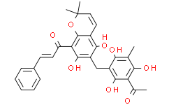
| Cas No.: | 82-08-6 |
| Chemical Name: | 2-Propen-1-one,1-[6-[(3-acetyl-2,4,6-trihydroxy-5-methylphenyl)methyl]-5,7-dihydroxy-2,2-dimethyl-2H-1-benzopyran-8-yl]-3-phenyl-,(2E)- |
| Synonyms: | 2-Propen-1-one,1-[6-[(3-acetyl-2,4,6-trihydroxy-5-methylphenyl)methyl]-5,7-dihydroxy-2,2-dimethyl-2H-1-benzopyran-8-yl]-3-phenyl-,(2E)-;2-Propen-1-one,1-[6-[(3-acetyl-2,4,6-trihydroxy-5-methylphenyl)methyl]-5,7-dihydroxy-2,2-dimethyl-2H-1-benzopyran-8-yl]-3-phe;pGlu-Ala-OH;Rottlerin;ROTTLERIN(RG);(E)-1-[6-[(3-acetyl-2,4,6-trihydroxy-5-methylphenyl)methyl]-5,7-dihydroxy-2,2-dimethylchromen-8-yl]-3-phenylprop-2-en-1-one;Kamalin;Mallotoxin |
| SMILES: | CC(C1=C(O)C(C)=C(O)C(CC2C(O)=C3C=CC(OC3=C(C(/C=C/C3C=CC=CC=3)=O)C=2O)(C)C)=C1O)=O |
| Formula: | C30H28O8 |
| M.Wt: | 516.538529396057 |
| Purity: | >98% |
| Sotrage: | 2 years -20°C Powder, 2 weeks 4°C in DMSO, 6 months -80°C in DMSO |
| Description: | Rottlerin is a specific PKC inhibitor, with IC50 values for PKCδ of 3-6 μM, PKCα,β,γ of 30-42 μM, PKCε,η,ζ of 80-100 μM. |
| Target: | PKCδ:3 μM (IC50, Porcine spleen) PKCα:30 μM (IC50, baculovirus-infected Sf9 insect cells) PKCγ:40 μM (IC50, baculovirus-infected Sf9 insect cells) PKCβ:42 μM (IC50, baculovirus-infected Sf9 insect cells) PKCη:82 μM (IC50, baculovirus-infected Sf9 insect cells) PKCζ:100 μM (IC50, baculovirus-infected Sf9 insect cells) PKCε:100 μM (IC50, baculovirus-infected Sf9 insect cells) CaM kinase III:5.3 μM (IC50, EF-2 kinase activity in cytosol of murine pancreas) CKII:30 μM (IC50, holoenzyme expressed in E.coli) PKA:78 μM (IC50, catalytic subunit from porcine heart) |
| In Vitro: | Rottlerin is a powerful inhibitor of the Ca2+-unresponsive PKCδ with an IC50 of 3 μM for the enzyme from porcine spleen and 6 μM for the recombinant enzyme from baculovirus-infected Sf9 insect cells. Other PKC isoenzymes provestobe at least one order of magnitude less sensitive for Rottlerin. The IC50 values for the different PKC isoenzymes increased in the following order: δ<α, β, γ<η, ζ, ε[1]. Rottlerin inhibits cell proliferation in a dose-dependent manner. The combination of both inhibitors (Sorafenib and Rottlerin) is substantially more effective than either single agent and produces a significant decrease in glioma cell survival. The cytotoxic effect of Rottlerin and Sorafenib is further confirmed using a clonogenic assay. There is a dose-dependent decrease in colony forming ability due to Rottlerin and Sorafenib, when administered independently, with the latter having activity at concentrations above 5 μM. In addition, there is striking potentiation of efficacy when the two agents are administered in combination[2]. |
| Cell Assay: | Cells (5×10³/well) are plated in 96-well microtiter plates in 100 μL of growth medium, and after overnight attachment, are exposed for 3 days to a range of concentrations of sorafenib and Rottlerin (0, 0.1, 0.2, 0.4, 0.8, 1.5, 3, 6, 12, 25, 50 μM), alone and in combination. Control cells receive vehicle alone (DMSO). After the treatment, cells are washed, and the number of viable cells is determined using a colorimetric cell proliferation assay. To perform the assay, 20 μL of MTS/phenazine methosulfate solution is added to each well, and after 1 h of incubation at 37°C in a humidified 5% CO2 atmosphere, absorbance is measured at 490 nm in a microplate reader. To directly assess cellular toxicity, 2.5×10⁵ cells are seeded in 60-mm Petri dishes and treated with selected concentrations of inhibitors or vehicle[2]. |
| References: | [1]. Gschwendt M, et al. Rottlerin, a novel protein kinase inhibitor. Biochem Biophys Res Commun. 1994 Feb 28;199(1):93-8. [2]. Jane EP, et al. Coadministration of sorafenib with rottlerin potently inhibits cell proliferation and migration in human malignant glioma cells. J Pharmacol Exp Ther. 2006 Dec;319(3):1070-80. |

 DC Chemicals' products qualify for U.S. tariff exemptions. We guarantee no price increases due to customs duties and maintain stable supply, continuing to deliver reliable research solutions to our American clients.


