| Description: |
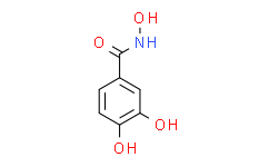
Didox is a synthetic ribonucleotide reductase (RR) inhibitor. |
| In Vitro: |
Didox suppresses LPS-induced mRNA levels of iNOS, IL-6, IL-1, TNF-α, NF-κβ (p65), and p38-α, after 24 h of treatment. Treatment with Didox also suppresses the secretion of nitric oxide (NO), IL-6, and IL-10. Using mitochondrial dehydrogenase activity as a measure of cytotoxicity, the effects of Didox on cellular respiration in RAW264.7 are examined over a range of concentrations for 24 h. Cells exposures to 200 μM and below Didox, with and without LPS, do not exhibit significant cellular toxicity[1]. Didox is active against all human and murine acute myeloid leukemia (AML) lines tested with IC50 values in the low micromolar range (mean IC50 37 µM [range 25.89-52.70 µM])[2]. |
| Cell Assay: |
RAW264.7 macrophages are treated with Didox alone, with 0.1 μg/mL LPS, or the two in combination. Cellular respiration, as an indication of cytotoxicity, is measured by the MTT assay, which quantifies mitochondrial dehydrogenase activity. Macrophages are plated into 96 well Costar plates at 105 cells per well in 100 μL of DMEM media. After 4 h of incubation at 37°C for adherence, compounds and DMSO carrier control (0.01% final) are added in triplicate over serial dilutions beginning with 200 μM per well in a total volume of 200 μL, and the plates incubated for 24 h. Four h before termination of the assay, each well receives 20 μL of a 5 mg/mL MTT solution in un-supplemented DMEM. After centrifugation, the supernatant for each well is discarded and cells containing reduced MTT are solubilized with 100 μL of acidified isopropanol (4 mM HCl, 0.1% NP-40 in isopropanol). Following a brief period of shaking, the optical density (O.D.) for each well is recorded at 550 nm. Each experiment is repeated three times and the data averaged from each triplicate, then expressed as percentage of the control O.D. values for each experiment[1]. |
| Animal Administration: |
Mice[2] Luciferase-tagged leukemia cells are transplanted into 8- week old, sublethally irradiated (4.5 Gy) C57Bl/6 mice by tail vein injection of 1.0×10⁶ cells per mouse. Mice are injected with 150 mg/kg D-Luciferin, anesthetised with Isoflurane, and imaged using the IVIS 100 imaging system. Mice begin treatment with Didox upon detection of clear signal. The animals are treated with daily administrations of Didox at 425 mg/kg Didox by intraperitoneal injection (IP) for 5 days. Control animals receive 5% dextrose water by IP injection. Repeat imaging is performed on the day following the final treatment[2]. |
| References: |
[1]. Matsebatlela TM, et al. 3,4-Dihydroxy-benzohydroxamic acid (Didox) suppresses pro-inflammatory profiles and oxidative stress in TLR4-activated RAW264.7 murine macrophages. Chem Biol Interact. 2015 May 25;233:95-105.
[2]. Cook GJ, et al. The efficacy of the ribonucleotide reductase inhibitor Didox in preclinical models of AML. PLoS One. 2014 Nov 17;9(11):e112619. |


